File: Canine lymphoma 1.JPG
Original file (1,192 × 885 pixels, file size: 124 KB, MIME type: image/jpeg)
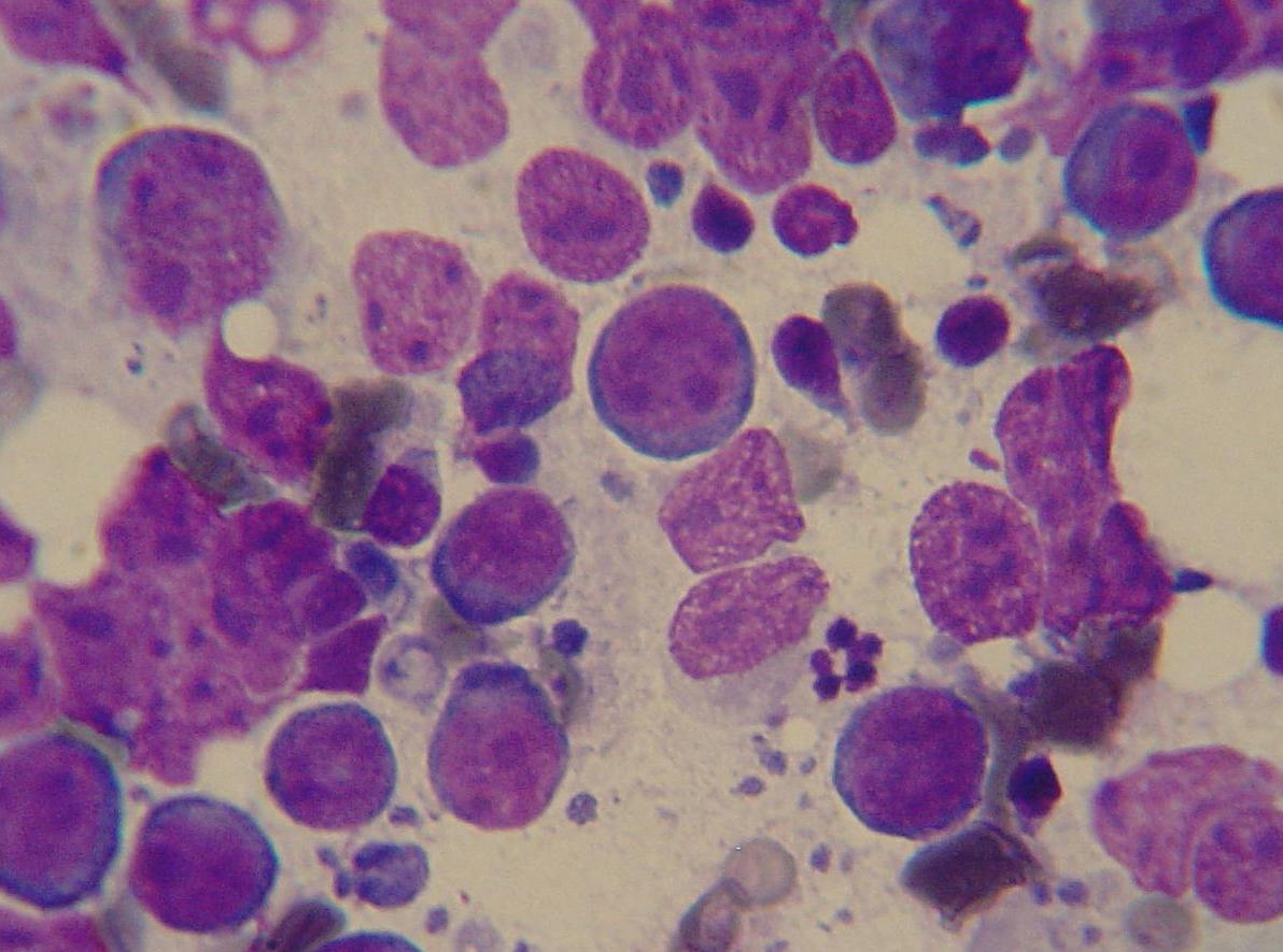
Cytology from a needle aspiration biopsy of a lymph node of a dog with lymphoma. The predominant cells are lymphoblasts. Slide was stained with a modified Wright's stain. Photo taken through a microscope at 100x by Joel Mills on September 17, 2005. Photo cropped on June 25, 2006.
Licensing
I, the author of this work, hereby publish it under the following licenses: Permission is granted to copy, distribute and/or modify this document under the terms of the GNU Free Documentation License, Version 1.2 or any later version published by the Free Software Foundation; with no Invariant Sections, no Front-Cover Texts, and no Back-Cover Texts. Subject to disclaimers.
This file is licensed under the Creative Commons Attribution ShareAlike license versions 2.5, 2.0, and 1.0.
You may select the license of your choice.
File history
Click on a date/time to view the file as it appeared at that time.
| Date/Time | Thumbnail | Dimensions | User | Comment | |
|---|---|---|---|---|---|
| current | 20:56, 10 July 2007 | | 1,192 × 885 (124 KB) | Rick Swarts (talk | contribs) | [http://en.wikipedia.org/wiki/Image:Canine_lymphoma_1.JPG source and rights] |
You cannot overwrite this file.
File usage
The following page uses this file: